Perforin-1
Mammalian protein found in Homo sapiens
| PRF1 | |||||||||||||||||||||||||||||||||||||||||||||||||||
|---|---|---|---|---|---|---|---|---|---|---|---|---|---|---|---|---|---|---|---|---|---|---|---|---|---|---|---|---|---|---|---|---|---|---|---|---|---|---|---|---|---|---|---|---|---|---|---|---|---|---|---|
 | |||||||||||||||||||||||||||||||||||||||||||||||||||
| Identifiers | |||||||||||||||||||||||||||||||||||||||||||||||||||
| Aliases | PRF1, FLH2, HPLH2, P1, PFN1, PFP, perforin 1 | ||||||||||||||||||||||||||||||||||||||||||||||||||
| External IDs | OMIM: 170280; MGI: 97551; HomoloGene: 3698; GeneCards: PRF1; OMA:PRF1 - orthologs | ||||||||||||||||||||||||||||||||||||||||||||||||||
| |||||||||||||||||||||||||||||||||||||||||||||||||||
| |||||||||||||||||||||||||||||||||||||||||||||||||||
| |||||||||||||||||||||||||||||||||||||||||||||||||||
| |||||||||||||||||||||||||||||||||||||||||||||||||||
| |||||||||||||||||||||||||||||||||||||||||||||||||||
| Wikidata | |||||||||||||||||||||||||||||||||||||||||||||||||||
| |||||||||||||||||||||||||||||||||||||||||||||||||||


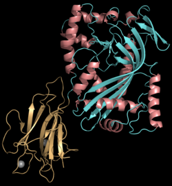
Perforin-1 Perforin (PRF), encoded by the PRF1 gene, is a pore-forming toxic protein housed in the secretory granules of cytotoxic T lymphocytes (CTLs) and natural killer (NK) cells. Together, these cells are known as cytotoxic lymphocytes (CLs). [5]
Discovery
Perforin was initially discovered in 1983 and subsequently cloned from an expression library in 1988 using anti-complement C9 antibody cross-reactivity. A sequence comparison showed a notable resemblance between the two proteins in a specific central region, termed the 'membrane attack complex/perforin' (MACPF) domain. [6]
Structure and Function
Purifying perforin is challenging due to its tendency to lose activity and stability in solution, and only recently has a recombinant form been successfully produced.[7]
Perforin is a pore forming cytolytic protein found in the granules of cytotoxic T lymphocytes (CTLs) and natural killer cells (NK cells). Upon degranulation, perforin molecules translocate to the target cell with the help of calreticulin, which works as a chaperone protein to prevent perforin from degrading. Perforin then binds to the target cell's plasma membrane via membrane phospholipids while phosphatidylcholine binds calcium ions to increase perforin's affinity to the membrane.[8] Perforin oligomerises in a Ca2+ dependent manner to form pores on the target cell. The pore formed allows for the passive diffusion of a family of pro-apoptotic proteases, known as the granzymes, into the target cell.[9] The lytic membrane-inserting part of perforin is the MACPF domain.[10] This region shares homology with cholesterol-dependent cytolysins from Gram-positive bacteria.[11]
The initial concept of a plasma membrane pore model was challenged when research revealed that granzyme B could undergo endocytosis independently of perforin. Moreover, experiments demonstrated that apoptosis could be induced by adding perforin to washed cells that had already endocytosed granzyme B, even in the absence of perforin during the endocytosis process. In light of these findings, Froelich and colleagues suggested that perforin's action might not occur at the plasma membrane as previously thought, but rather at the endosomal membrane.[12] They proposed that perforin likely facilitates the release of granzymes from endosomes by forming pores in the endosomal membrane.[7]
Perforin has structural and functional similarities to complement component 9 (C9). Like C9, this protein creates transmembrane tubules and is capable of lysing non-specifically a variety of target cells. This protein is one of the main cytolytic proteins of cytolytic granules, and it is known to be a key effector molecule for T-cell- and natural killer-cell-mediated cytolysis.[13] Perforin is thought to act by creating holes in the plasma membrane which triggers an influx of calcium and initiates membrane repair mechanisms. These repair mechanisms bring perforin and granzymes into early endosomes.[14]
Clinical Significance
Acute presentations of perforinopathy — FHL
Around the turn of the 21st century, it was recognized that a total loss of perforin activity leads to a severe, fatal autosomal recessive immunoregulatory disorder in infants, known as familial hemophagocytic lymphohistiocytosis (FHL), typically appearing before 12 months of age. This condition can only be treated effectively with a bone marrow transplant from a non-related donor.[5]
The pathogenesis involves a sequence of downstream events resulting from the inability of NK cells and CTLs to present functional perforin, thereby failing to kill target cells. Infants affected by this condition typically show symptoms of “classic” familial hemophagocytic lymphohistiocytosis (FHL) and meet most or all of the criteria outlined in HLH-2004. Diagnosis is confirmed by reduced NK cell cytotoxicity, as healthy NK cells normally exhibit constitutive, pathogen-independent cytotoxicity, and by identifying mutations in genes such as PRF1, UNC13D, STX11, and STXBP2.[5]
Sub-acute presentations of perforinopathy
Sub-acute perforinopathies encompass a diverse array of symptoms, all stemming from a partial ("sub-total") reduction in cytotoxic lymphocyte (CL) activity due to bi-allelic mutations in one of the four previously mentioned genes. In contrast to the acute form, diagnosing sub-acute perforinopathies can be challenging due to their typically milder and more sporadic clinical manifestations, an intermittent disease course, variability in onset age, and their frequent positive response to non-specific immune-suppressive or immune-ablative treatments.[5]
Chronic presentations of perforinopathy
Chronic perforinopathies are regarded as a range of immune-related conditions resulting from monoallelic mutations in genes linked to familial hemophagocytic lymphohistiocytosis (FHL). These conditions typically manifest differently from classic FHL and may include conditions like blood cancers and macrophage activation syndrome, particularly in patients with juvenile rheumatoid arthritis. Onset of symptoms usually occurs after the age of 5. Moreover, some research suggests a correlation between variations in the PRF1 gene and the outcome of allogeneic bone marrow transplantation. However, these associations remain controversial, with studies disproving the connection outweighing those supporting it.[5]
Interactions
Perforin has been shown to interact with calreticulin.[15]
See also
References
- ^ a b c GRCh38: Ensembl release 89: ENSG00000180644 – Ensembl, May 2017
- ^ a b c GRCm38: Ensembl release 89: ENSMUSG00000037202 – Ensembl, May 2017
- ^ "Human PubMed Reference:". National Center for Biotechnology Information, U.S. National Library of Medicine.
- ^ "Mouse PubMed Reference:". National Center for Biotechnology Information, U.S. National Library of Medicine.
- ^ a b c d e Voskoboinik, Ilia; Trapani, Joseph A. (2013). "Perforinopathy: A Spectrum of Human Immune Disease Caused by Defective Perforin Delivery or Function". Frontiers in Immunology. 4: 441. doi:10.3389/fimmu.2013.00441. ISSN 1664-3224. PMC 3860100. PMID 24376445.
- ^ Brennan, A J; Chia, J; Trapani, J A; Voskoboinik, I (April 2010). "Perforin deficiency and susceptibility to cancer". Cell Death & Differentiation. 17 (4): 607–615. doi:10.1038/cdd.2009.212. ISSN 1350-9047. PMID 20075937.
- ^ a b Pipkin, Matthew E; Lieberman, Judy (2007-06-01). "Delivering the kiss of death: progress on understanding how perforin works". Current Opinion in Immunology. Lymphocyte activation/Lymphocyte effector functions. 19 (3): 301–308. doi:10.1016/j.coi.2007.04.011. ISSN 0952-7915. PMID 17433871.
- ^ Osińska, Iwona et al. “Perforin: an important player in immune response.” Central-European journal of immunology vol. 39,1 (2014): 109-15. doi:10.5114/ceji.2014.42135
- ^ Trapani JA (1996). "Target cell apoptosis induced by cytotoxic T cells and natural killer cells involves synergy between the pore-forming protein, perforin, and the serine protease, granzyme B". Australian and New Zealand Journal of Medicine. 25 (6): 793–9. doi:10.1111/j.1445-5994.1995.tb02883.x. PMID 8770355.
- ^ Tschopp J; Masson D; Stanley KK (1986). "Structural/functional similarity between proteins involved in complement- and cytotoxic T-lymphocyte-mediated cytolysis". Nature. 322 (6082): 831–4. Bibcode:1986Natur.322..831T. doi:10.1038/322831a0. PMID 2427956. S2CID 4330219.
- ^ Rosado CJ, Buckle AM, Law RH, Butcher RE, Kan WT, Bird CH, Ung K, Browne KA, Baran K, Bashtannyk-Puhalovich TA, Faux NG, Wong W, Porter CJ, Pike RN, Ellisdon AM, Pearce MC, Bottomley SP, Emsley J, Smith AI, Rossjohn J, Hartland EL, Voskoboinik I, Trapani JA, Bird PI, Dunstone MA, Whisstock JC (2007). "A common fold mediates vertebrate defense and bacterial attack". Science. 317 (5844): 1548–51. Bibcode:2007Sci...317.1548R. doi:10.1126/science.1144706. PMID 17717151. S2CID 20372720.
- ^ Froelich, Christopher J.; Orth, Kim; Turbov, Jane; Seth, Prem; Gottlieb, Roberta; Babior, Bernard; Shah, Girish M.; Bleackley, R. Christopher; Dixit, Vishva M.; Hanna, William (November 1996). "New Paradigm for Lymphocyte Granule-mediated Cytotoxicity". Journal of Biological Chemistry. 271 (46): 29073–29079. doi:10.1074/jbc.271.46.29073. ISSN 0021-9258. PMID 8910561.
- ^ "Entrez Gene: PRF1 perforin 1 (pore forming protein)".
- ^ Thiery J, Keefe D, Boulant S, Boucrot E, Walch M, Martinvalet D, Goping IS, Bleackley RC, Kirchhausen T, Lieberman J (2011). "Perforin pores in the endosomal membrane trigger the release of endocytosed granzyme B into the cytosol of target cells". Nat. Immunol. 12 (8): 770–7. doi:10.1038/ni.2050. PMC 3140544. PMID 21685908.
- ^ Andrin C, Pinkoski MJ, Burns K, et al. (July 1998). "Interaction between a Ca2+-binding protein calreticulin and perforin, a component of the cytotoxic T-cell granules". Biochemistry. 37 (29): 10386–94. doi:10.1021/bi980595z. PMID 9671507.
Further reading
- Trapani JA (1996). "Target cell apoptosis induced by cytotoxic T cells and natural killer cells involves synergy between the pore-forming protein, perforin, and the serine protease, granzyme B". Australian and New Zealand Journal of Medicine. 25 (6): 793–9. doi:10.1111/j.1445-5994.1995.tb02883.x. PMID 8770355.
- Peitsch MC, Amiguet P, Guy R, et al. (1990). "Localization and molecular modelling of the membrane-inserted domain of the ninth component of human complement and perforin". Mol. Immunol. 27 (7): 589–602. doi:10.1016/0161-5890(90)90001-G. PMID 2395434.
- Young JD, Hengartner H, Podack ER, Cohn ZA (1986). "Purification and characterization of a cytolytic pore-forming protein from granules of cloned lymphocytes with natural killer activity". Cell. 44 (6): 849–59. doi:10.1016/0092-8674(86)90007-3. PMID 2420467. S2CID 30182487.
- Young JD, Cohn ZA, Podack ER (1986). "The ninth component of complement and the pore-forming protein (perforin 1) from cytotoxic T cells: structural, immunological, and functional similarities". Science. 233 (4760): 184–90. doi:10.1126/science.2425429. PMID 2425429.
- Lichtenheld MG, Podack ER (1990). "Structure of the human perforin gene. A simple gene organization with interesting potential regulatory sequences". J. Immunol. 143 (12): 4267–74. doi:10.4049/jimmunol.143.12.4267. PMID 2480391. S2CID 8326644.
- Shinkai Y, Takio K, Okumura K (1988). "Homology of perforin to the ninth component of complement (C9)". Nature. 334 (6182): 525–7. Bibcode:1988Natur.334..525S. doi:10.1038/334525a0. PMID 3261391. S2CID 4348928.
- Lichtenheld MG, Olsen KJ, Lu P, et al. (1988). "Structure and function of human perforin". Nature. 335 (6189): 448–51. Bibcode:1988Natur.335..448L. doi:10.1038/335448a0. PMID 3419519. S2CID 4359028.
- Goebel WS, Schloemer RH, Brahmi Z (1996). "Target cell-induced perforin mRNA turnover in NK3.3 cells is mediated by multiple elements within the mRNA coding region". Mol. Immunol. 33 (4–5): 341–9. doi:10.1016/0161-5890(95)00155-7. PMID 8676885.
- Nöske K, Bilzer T, Planz O, Stitz L (1998). "Virus-Specific CD4+ T Cells Eliminate Borna Disease Virus from the Brain via Induction of Cytotoxic CD8+ T Cells". J. Virol. 72 (5): 4387–95. doi:10.1128/JVI.72.5.4387-4395.1998. PMC 109669. PMID 9557729.
- Andrin C, Pinkoski MJ, Burns K, et al. (1998). "Interaction between a Ca2+-binding protein calreticulin and perforin, a component of the cytotoxic T-cell granules". Biochemistry. 37 (29): 10386–94. doi:10.1021/bi980595z. PMID 9671507.
- Yu CR, Ortaldo JR, Curiel RE, et al. (1999). "Role of a STAT binding site in the regulation of the human perforin promoter". J. Immunol. 162 (5): 2785–90. doi:10.4049/jimmunol.162.5.2785. PMID 10072525. S2CID 26096007.
- Stepp SE, Dufourcq-Lagelouse R, Le Deist F, et al. (1999). "Perforin gene defects in familial hemophagocytic lymphohistiocytosis". Science. 286 (5446): 1957–9. doi:10.1126/science.286.5446.1957. PMID 10583959.
- Takahashi T, Nieda M, Koezuka Y, et al. (2000). "Analysis of human V alpha 24+ CD4+ NKT cells activated by alpha-glycosylceramide-pulsed monocyte-derived dendritic cells". J. Immunol. 164 (9): 4458–64. doi:10.4049/jimmunol.164.9.4458. PMID 10779745.
- Badovinac VP, Tvinnereim AR, Harty JT (2000). "Regulation of antigen-specific CD8+ T cell homeostasis by perforin and interferon-gamma". Science. 290 (5495): 1354–8. Bibcode:2000Sci...290.1354B. doi:10.1126/science.290.5495.1354. PMID 11082062.
- Göransdotter Ericson K, Fadeel B, Nilsson-Ardnor S, et al. (2001). "Spectrum of Perforin Gene Mutations in Familial Hemophagocytic Lymphohistiocytosis". Am. J. Hum. Genet. 68 (3): 590–7. doi:10.1086/318796. PMC 1274472. PMID 11179007.
- Clementi R, zur Stadt U, Savoldi G, et al. (2002). "Six novel mutations in the PRF1 gene in children with haemophagocytic lymphohistiocytosis". J. Med. Genet. 38 (9): 643–6. doi:10.1136/jmg.38.9.643. PMC 1734943. PMID 11565555.
- Ambach A, Bonnekoh B, Gollnick H (2001). "Perforin granule release from cytotoxic lymphocytes ex vivo is inhibited by ciclosporin but not by methotrexate". Skin Pharmacol. Appl. Skin Physiol. 14 (5): 249–60. doi:10.1159/000056355 (inactive 2024-06-11). PMID 11586066. S2CID 30142804.
{{cite journal}}: CS1 maint: DOI inactive as of June 2024 (link)
External links
- perforin at the U.S. National Library of Medicine Medical Subject Headings (MeSH)
Perforin-1 at NLM Genetics Home Reference


